Courtesy of Intermountain Medical Imaging, Boise, Idaho.
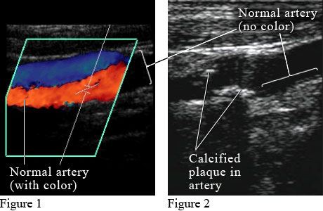
Figure 1 shows a color Doppler ultrasound picture of blood flowing through a normal artery (red) and vein (blue). Figure 2 shows an ultrasound picture of an artery narrowed by the buildup of calcium and fat (cholesterol) in the inner lining of the artery, called plaque, which leads to "hardening of the arteries" (atherosclerosis).
Current as of: July 31, 2024
Author: Ignite Healthwise, LLC Staff
Clinical Review Board
All Healthwise education is reviewed by a team that includes physicians, nurses, advanced practitioners, registered dieticians, and other healthcare professionals.