Courtesy of Intermountain Medical Imaging, Boise, Idaho. All rights reserved.
An angiogram is an X-ray test done to take pictures of the blood flow in an artery or vein. During the test, you lie on a table and a dye is injected into a blood vessel in the groin or elbow. A special camera takes pictures of the dye as it moves through the blood vessels.
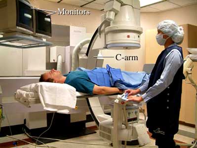
In this picture, the radiologist uses monitors and table controls to position the person before an angiogram. The large C-arm can turn to take X-ray pictures from many different angles.
Current as of: July 31, 2024
Author: Ignite Healthwise, LLC Staff
Clinical Review Board
All Healthwise education is reviewed by a team that includes physicians, nurses, advanced practitioners, registered dieticians, and other healthcare professionals.